Ultrasound probes are widely used medical devices that routinely contact intact skin or mucous membranes(e.g., endocavity probes). To prevent patient-to-patient transmission of microbes, probes are cleaned and, when indicated, subjected to high level disinfection (HLD). As probes age or are handled roughly, microfractures, microscopic cracks, scratches, or separations can develop in the probe’s external surface or protective layers.
The question for healthcare professionals and those tasked with patient safety is whether microfractures can cause ultrasound probes to harbor bacteria and other harmful soils after cleaning and high-level disinfection, why this matters, and what practical steps can reduce the risks.
What Are Microfractures and How Are They Caused?
Microfractures are tiny defects in an ultrasound probe’s outer materials, such as the acoustic lens, polymer membrane, or protective overmold.
They can be caused by:
- • Mechanical stress (drops, impact, repeated flexing).
- • Chemical degradation (incompatible disinfectants or repeated exposure).
- • Wear from repeated use and cleaning cycles.
- • Manufacturing defects or adhesive failures in multilayer assemblies.
- • Excessive exposure to heat beyond the probe manufacturer’s guidelines.
- • Incorrect materials used during a third-party repair.
- • Exposure to shortwave UV light for extended periods or repetitive exposure.
These defects can be so small that they are invisible to the naked eye, but large enough to create a protected microenvironment at the surface.
How Microfractures Can Harbor Microbes and Soils
- Protected niches: Microfractures can shelter organic soils (blood, mucus, gel residues) and microorganisms, physically blocking disinfectant access. Chemical agents rely on contact; if organic material or crevices prevent contact, microbes may survive.
- Facilitation of biofilms: Micro-crevices provide surfaces where microbes can adhere and begin forming biofilms. Biofilms are communities of microbes embedded in an extracellular matrix that can greatly increase tolerance to disinfectants compared with other microbes.
- Residual proteins and gel: Gel, blood, and proteinaceous soils can lodge in cracks. Organic matter both shields microbes and consumes disinfectant, reducing effective concentration.
Effectiveness of HLD in the Presence of Microfractures
High level disinfectants (e.g., glutaraldehyde, ortho phthalaldehyde, peracetic acid, and hydrogen peroxide formulations) are effective against a broad range of bacteria, mycobacteria, fungi, and viruses when used per manufacturer instructions (concentration, contact time, temperature, and prior cleaning). However, their effectiveness assumes adequate cleaning and direct contact with contaminated surfaces. Beyond HLD chemicals, consideration must be given to the effects on construction materials and the probes' plastics, polymers, and glues when UV light is used for high-level disinfection.
Problems That Can Arise When Ultrasound Probes Contain Microfractures
- • Disinfectant penetration into microscopic cracks can be incomplete, especially if soil or biofilm is present.
- • Micro-crevices are heavily shadowed and UV light is not able to penetrate.
- • Some pathogens (or pathogens in biofilms) demonstrate increased tolerance of disinfectants; non enveloped viruses and spores are inherently more resistant and may survive if contact is insufficient.
- • Several reports and studies1 have documented the detection of microbial DNA (including viruses) or viable organisms on probe surfaces after standard HLD, sometimes linked to surface damage or inadequate cleaning; while detection of DNA does not always equal infectious risk, the presence of viable microbes or biofilm raises concern.
Clinical Significance and Documented Risks
Actual documented infections directly attributable to microfractures in probes are relatively uncommon, but case reports and surveillance studies have highlighted lapses in infection prevention linked to probe surface defects, inadequate cleaning, or breaches of probe-cover protocols. The risk depends on the procedure type: it is higher for probes that contact mucous membranes (endocavity or transrectal/transvaginal uses) than for external, superficial probes. Vulnerable patients (immunocompromised) and invasive procedures increase potential consequences.
How to Detect and Assess Microfractures in Ultrasound Probes
- • Visual inspection: viewing a probe under good lighting and magnification can reveal scratches, cracks, or delamination.
- • Tactile inspection and dye tests: some centers use dyes or methylene blue to find cracks in probes.
- • Manufacturer-recommended leak testing: many probe manufacturers provide specific leak test kits or procedures to detect breaches in the acoustic lens or internal seals.
- • Regular preventive maintenance and electrical/functional testing are also advised; once microfractures are detected, the device should be evaluated by the manufacturer.
Mitigation Strategies and Best Practices
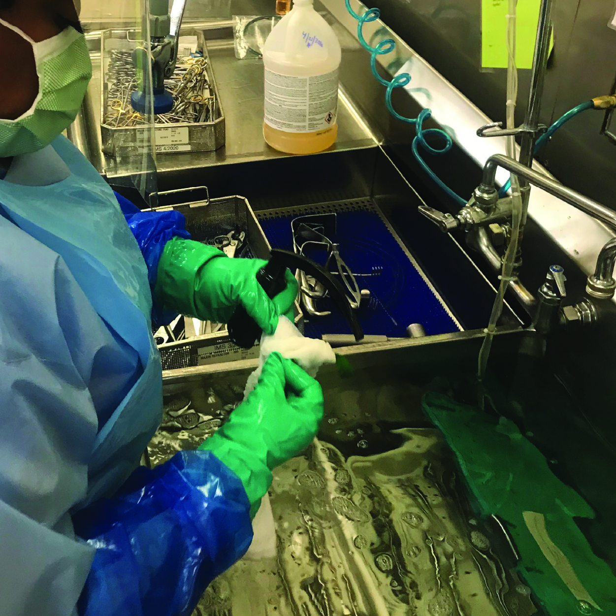
Point-of-Use cleaning, bedside cleaning, and pre-cleaning: Immediate cleaning to remove large amounts of debris, helping with cleaning and preventing debris from drying on the medical device.
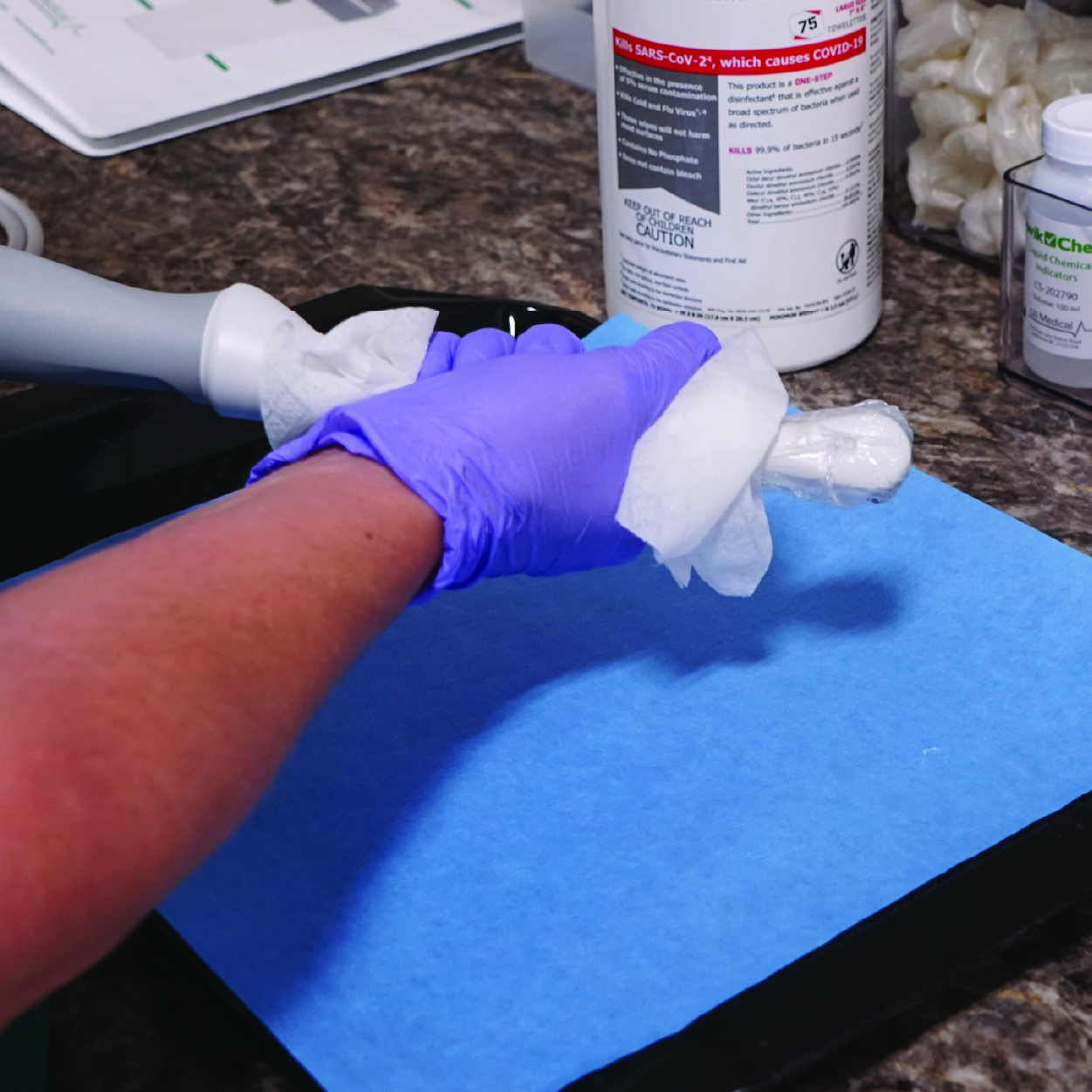
Cleaning: Thorough cleaning (automated or manual) to remove gels and organic soils before disinfection with liquid or UV light is critical because it enables disinfectant access. Use enzymatic detergents when indicated by the manufacturer’s IFU.
Disinfection: Follow manufacturer instructions. Use only approved disinfectants, concentrations, and contact times. Some disinfectants may degrade materials and accelerate microfracturing — consult the manufacturer's material compatibility guidelines.
Routine inspection and leak testing: Implement scheduled visual and leak tests, with immediate action (repair or decommission) when defects are found. Some testing must be performed between every procedure.
Use of probe covers/sheaths: For endocavity procedures, single use probe covers are required to reduce contamination. Remember that covers can fail (micro-perforations), so they are an adjunct, not a substitute for cleaning and high-level disinfection.
Sterile gel for invasive procedures: Use sterile coupling gel inside covers and for invasive procedures to reduce the introduction of microbes.
Repair or replace damaged probes: Once microfractures or delamination are detected, follow manufacturer guidance. Many recommend removing the probe from service for repair or replacement, as repair options are limited and field repairs may be unsafe.
Staff training and policies: Proper technique for all steps of reprocessing, such as cleaning, disinfecting, rinsing, handling, and storage, reduces mechanical damage and contamination risks. Maintain documentation of maintenance, testing, and incidents.
Key Takeaways
Microfractures in ultrasound probes create protected niches that can shelter organic soils and microbes, potentially reducing the effectiveness of high level disinfection. While liquid chemical HLD is highly effective when surfaces are intact and properly cleaned, microfractures compromise contact and can allow microbes to survive or persist, potentially including organisms in biofilms. The risk of resulting patient infection is context dependent (type of procedure, organism, host susceptibility), but prudent infection prevention requires preventive measures: thorough cleaning, routine inspection and leak testing, use of covers, sterile gel for invasive procedures, adherence to manufacturer instructions, and prompt removal/repair of damaged probes. Continued research, robust device design standards, and strict operational policies are essential to minimize this preventable risk.